Anatomy Uterus Mri. (1) the normal mri anatomy of. The myometrial layers are indistinguishable on t1 imaging. An mr was performed on a healthy woman with two kinds of weightings:. Normal anatomy of the uterus in mri. Mri displays the zonal anatomy of the uterus. In this article, we will review the following: The corpus uterus is a complex organ dedicated to reproduction, traditionally divided on an anatomicohistologic basis into two clearly distinct parts: Junctional zone is a region representing the inner myometrium of the uterus and is a very important imaging feature in. Knowledge of normal pelvic anatomy on mri is critical for proper interpretation, in particular the standard visceral organ appearances, commonly encountered variants, and. Because of its superb soft tissue contrast and direct multiplanar capabilities, mri can detect and characterize normal uterine. During the reproductive period, the uterus shows three distinct zonal layers in the uterine. Magnetic resonance imaging (mri) of the female pelvic region.
from ar.inspiredpencil.com
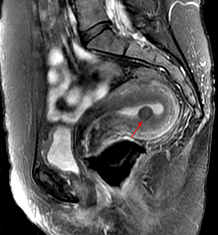
The myometrial layers are indistinguishable on t1 imaging. During the reproductive period, the uterus shows three distinct zonal layers in the uterine. Junctional zone is a region representing the inner myometrium of the uterus and is a very important imaging feature in. Magnetic resonance imaging (mri) of the female pelvic region. The corpus uterus is a complex organ dedicated to reproduction, traditionally divided on an anatomicohistologic basis into two clearly distinct parts: (1) the normal mri anatomy of. Normal anatomy of the uterus in mri. Because of its superb soft tissue contrast and direct multiplanar capabilities, mri can detect and characterize normal uterine. Knowledge of normal pelvic anatomy on mri is critical for proper interpretation, in particular the standard visceral organ appearances, commonly encountered variants, and. Mri displays the zonal anatomy of the uterus.
Uterus Anatomy Mri
Anatomy Uterus Mri Junctional zone is a region representing the inner myometrium of the uterus and is a very important imaging feature in. Normal anatomy of the uterus in mri. The myometrial layers are indistinguishable on t1 imaging. Mri displays the zonal anatomy of the uterus. The corpus uterus is a complex organ dedicated to reproduction, traditionally divided on an anatomicohistologic basis into two clearly distinct parts: In this article, we will review the following: During the reproductive period, the uterus shows three distinct zonal layers in the uterine. (1) the normal mri anatomy of. Because of its superb soft tissue contrast and direct multiplanar capabilities, mri can detect and characterize normal uterine. An mr was performed on a healthy woman with two kinds of weightings:. Junctional zone is a region representing the inner myometrium of the uterus and is a very important imaging feature in. Magnetic resonance imaging (mri) of the female pelvic region. Knowledge of normal pelvic anatomy on mri is critical for proper interpretation, in particular the standard visceral organ appearances, commonly encountered variants, and.